The Vector
Volume 6, Issue 6: November 2017
Editorial Team
Guohua Yi, PhD - Editor, The Vector
Phillip Doerfler, PhD - Associate Editor, The Vector
Melvin Rincon, MD, PhD - Junior Editor, The Vector
Inside This Issue
President's Message
Breaking Through
Society News
Public Policy
Industry News

President's Message
 Dear Colleagues,
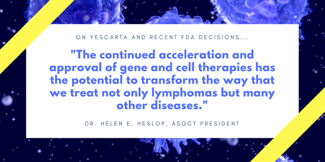
Last month I attended the 25th meeting of the European Society for Gene and Cell Therapy in Berlin which was an excellent meeting characterized by lots of opportunities for interactions and high quality food and wine. It was also very gratifying to hear about multiple approaches in different diseases that have been optimized by iterative bench to bedside testing and are now moving towards definitive clinical testing and licensure. During the meeting, the FDA granted approval of Yescarta (axicabtagene ciloleucel) to Kite Pharma, Inc. which is the second CD19 CAR T strategy approved this year by the FDA and the first to target adult patients with certain types of large B-cell lymphoma who have not responded to or who have relapsed after at least two other kinds of treatment. Both Yescarta and Kymriah approved for relapsed/refractory pediatric ALL offer a potentially curative option to patients with a very poor prognosis and are a major advance in the field. There a number of issues to be addressed though to ensure access to all patients who could benefit including development of new codes to cover the product and care episodes and evaluation of novel value based pricing models.
There was another major event for the field in early October, when the FDA advisory committee unanimously recommended approval for Spark Therapeutics gene therapy, Luxturna which treats inherited retinal diseases caused by defects in a gene known as RPE65. This is a step toward the FDA’s first approved gene therapy for an inherited disease. A final decision is expected by January 12, 2018.
One of the major strategic efforts of the society in the last year has been to upgrade the ASGCT website and after many months of effort on the part of the Board of Directors, Outreach & Communications Committee, and staff, I am pleased to announce that ASGCT has launched the new website. The new website provides a more member-friendly experience with vast improvements to the navigation, content, and aesthetic appeal and I encourage you to explore it and give any feedback to the ASGCT office.
In the coming months, please do not forget to renew your membership for 2018. Take a moment to visit the website to quickly and securely renew your membership. Members receive discounted registration rates for the 21st Annual Meeting, a subscription to the Molecular Therapy Family of Journals, access to the job bank, a variety of networking opportunities, and much more.
As we look ahead to 2018, there are many great opportunities to get involved in the 21st Annual Meeting. Abstract submission is now open! Please submit your abstract before the January 31st deadline. This is a great opportunity to present and discuss your research with your peers and leaders in the field. Associate Members who are the first and presenting author on an abstract will receive free Annual Meeting registration. Awards and travel grants are given to approximately 150 abstract submitters based on abstract scores. Additionally, ASGCT is accepting nominations for the 2018 Outstanding Achievement Award, Outstanding New Investigator Awards, and Sonia Skarlatos Public Service Award through December 15th. To learn more about each award and to review the criteria for each please visit award nominations page.
Breaking Through

Kole et al. Mol Ther. doi: 10.1016/j.ymthe.2017.09.007
Summary written by: Thierry Léveillard, Christo Kole and Laurence Klipfel
In the retina of vertebrates, the capture of photons of incident light, the initial step of visual perception by photoreceptor cells, results from the trans-isomerization of a derivative of vitamin A covalently linked to a seven-transmembrane, G protein–coupled receptor, the opsin molecule. Opsins are highly concentrated in a subcellular structure, the outer segment, made of multiple disks or of invaginations of lipid bilayers, in rod and cone photoreceptors, respectively. This cyto-architecture permits maximal light perception. The trans-isomerization of the vitamin A-derivative triggers a conformation change in the seven surrounding helixes of opsin and initiates the phototransduction cascade. This is facilitated by the optimal fluidity of the lipids of the outer segments that are rich in poly-unsaturated fatty acids (PUFA). The sophistication of this photon-capture system is the cause of its vulnerability. PUFA are prone to oxidation, either by reactive oxygen species produced by metabolic leakage of the mitochondrial respiratory chain or directly by photo-oxidation. The outer segment must constantly be renewed to keep the system operational.
Photoreceptor outer segments point in the direction of the retinal pigment epithelium (RPE), corresponding to the opposite direction to the incident light. RPE cells phagocytize daily 10% of the outer segments that are then renewed by synthesis in the photoreceptor inner segment. Glucose supply from blood circulation through the RPE is metabolized by photoreceptors through aerobic glycolysis to produce triglycerides entering in the composition of phospholipids of the outer segments. For this gigantic energetic task, rods assist metabolically cones. This ménage à trois (RPE-rods-cones) is reflected in the pathophysiology of retinal diseases. Retinitis pigmentosa (RP), the most prevalent Mendelian retinal degeneration, occurs when any of the 63 genes identified so far is mutated. Rods degenerate through apoptosis leading to night vision, but also leaving cones without metabolic assistance. The predominance of rods over cones, a contingence linked to the evolutionary history of the mammalian retina, results in the dysfunction of cones and ultimately to their death, the consequence is an irreversible blindness for RP patients. In primates, cones are concentrated at a specific point, called the fovea within the central region of the retina, the macula. This region that sustains most of our visual acuity is also affected in another and more frequent blinding disease, aged-related macular degeneration (AMD). RP genes have nothing in common with the 34 loci where AMD risk alleles are located indicating that the function of cones is harmed in these retinal diseases by distinct pathophysiological mechanisms that converge.
The genetic heterogeneity of RP and the genetic complexity of AMD were the key arguments for the development of therapeutic approaches that target common pathways that would alleviate the need for gene- and mutation-restricted cures to treat these threatening diseases. The restoration of metabolic and redox signaling between rods and cones by the products of the NXNL1 is one of such approaches for RP. Neovascularization is a clinical sign that is amenable to anti-VEGF therapy of AMD, but concerns only a minority of patients. More generally, oxidative damages accumulation with age and the chronic non-resolving inflammation are environmental risk factors. The production of cytotoxic byproducts of vitamin A close to RPE cells might participate in disease progression that is initiated in genetically predisposed people by local and abnormal activation of the complement cascade. Because the dysfunction and progressive destruction of the RPE are considered as instrumental, replacing the affected RPE by transplantation of unaffected one has been envisioned for years as a potential treatment of AMD.
In medical practice, cell transplantation requires the production of healthy RPE cells. Consequently, the expected therapeutic benefit on AMD will depend on the ability to produce RPE cells that must be amplified in vitro before transplantation. Our study reveals that the expansion of primary pig RPE cells is accompanied by the reduction of the expression of messenger RNAs of 27 out of 37 genes involved in RPE function, designated later as RPE markers. Among these genes, two transcription factors of the homeobox family, OTX2 and CRX were selected as candidate upstream regulators of a gene network preventing the down-regulation of the RPE markers. OTX2 was further studied since it was shown to regulate CRX expression and because its inactivation in mature RPE of the mouse leads to retinal dysfunction. We demonstrated the ability of OTX2 to restore the expression of 10 of these RPE markers after the infection of pig primary RPE cells with a recombinant adeno-associated vector (AAV) serotype 1. A direct effect of OTX2 on the activity of promoter of these RPE markers was demonstrated in a subset of four genes. To ensure that the observed phenomenon was not restricted to the studied species, we performed a similar analysis with human induced pluripotent (iPS) cells differentiated into RPE and confirm that OTX2 enhances the expression of CRX, but also of the potassium voltage-gated channel KCNJ13 and the lactate transporter SLC16A8.
Because the retina of rodents do not have a macula, we pursued our study in vivo with the Royal College of Surgeon (RCS) rat, a spontaneous model of a recessive form of RP with a mutation in the gene Mertk, expressed specifically by RPE cells and involved in photoreceptor outer segments phagocytosis. Pig primary RPE cells were cultured for one week while infected with AAV2/1-OTX2 or AAV2/1-GFP as negative control. Those cells (50,000 cells/eye) were then injected in the subretinal space of the RCS rat aged 18 days. In this model of RP, both rods and cones degenerate, starting from day 19. The function of photoreceptors was measured using electroretinograhy (ERG). The rod response was maintained in the grafted animals as compared to the unoperated ones, in accordance to previous observations. More importantly, the over-expression of OTX2 in the transplanted cells improve significantly rod function, but also that of cones. The transplantation of RPE-GFP cells increases cone response by 186% and that of RPE-OTX2 cells by 305%. OTX2 increases the effect of RPE transplantation by 164% compared to GFP. Given the average loss of cone vision of about 4% per year for RP patients, 164% would refer to 41 years of maintenance of central vision, a medically significant situation.
The preservation of rod function is correlated with a reduction of the death of rod photoreceptors as scored by optic coherence tomography (OCT). The protection of rods is likely the source of the maintenance of cone function through the NXNL1 metabolic signaling. The mechanism of action of OTX2 in those experimental conditions is not completely elucidated. The fact that rod preservation was observed at distance from the transplanted cells indicates that the visual benefit does not entirely rely on the restoration of photoreceptor outer segments phagocytosis that is defective in the RCS rat. Our analysis supports the presence of unidentified protective factors secreted by RPE cells transduced with OTX2. We also highlight that the up-regulation of proteins regulating metabolites transport and transmembrane ion gradients could also produce the non cell-autonomous protection of rods.
The maintenance of a proper differentiation status of the grafted cells by OTX2 is relevant for the treatment of AMD by iPS cell transplantation. In that perspective, the lactate transporter SLC16A8 whose expression in the RPE is directly or indirectly regulated by OTX2 carries a risk allele for AMD.
Programmable base editing of A•T to G•C in genomic DNA without DNA cleavage
N. Gaudelli et al., “Programmable base editing of A•T to G•C in genomic DNA without DNA cleavage,” Nature, doi:10.1038/nature24644, 2017.
Summarized Nicole M. Gaudelli and David R. Liu
In this work we report the development of a new base editor that enables the clean conversion of an A•T base pair into a G•C base pair without breaking the DNA backbone (in contrast with current CRISPR-Cas9-mediated HDR technologies). Last year, our group created a related base editor, BE3, which converts C•G base pairs to T•A through the formation of a uracil intermediate. BE3, and its improved variant BE4 reported earlier this year, consists of a cytosine deaminase (APOBEC1), which operates efficiently on ssDNA but not dsDNA, to the N-terminal portion of a catalytically impaired nickase mutant of the S. pyogenes Cas9 endonuclease (nCas9). The nCas9 potion of BE3/BE4 binds to a target DNA sequence in a guide RNA-programmed manner, and creates a DNA-RNA heteroduplex (R loop) containing an unpaired strand of the target genomic dsDNA. Within the small ssDNA bubble in this R loop, cytosines are suitably positioned as substrates for APOBEC-mediated hydrolytic deamination. This deamination process chemically converts cytosine into uracil, a base recognized as thymine (T) by DNA and RNA polymerases. Critically, the nCas9 portion of the BE3/BE4 base editors cuts only the strand of DNA that is not deaminated. This key feature of the BE3/BE4 base editors induces the cell to repair the unedited (G-containing) strand using the U-containing strand as a template. The net result is replacement of the target C•G base pair with a T•A base pair. In order to avoid interference from cellular uracil DNA glycosylase, which initiates base excision repair of U that would derail the base editing process, BE3 and BE4 also contain one and two copies, respectively, of uracil glycosylase inhibitor (UGI), which impede uracil excision.
Similar to BE3 and BE4, the adenine base editor (ABE) described in this new work also uses a deaminase fused to the N-terminus of nCas9. The deaminase used in ABE catalyzes the hydrolytic deamination of adenine (A), forming inosine (I), which is read as guanine (G) by DNA and RNA polymerases. However, in contrast with the abundance of naturally occurring enzymes that convert C to U in DNA, no enzymes are known to catalyze A to I formation in DNA. Although several enzymes are known to catalyze A to I formation in RNA, we showed that when fused to nCas9, these RNA adenine deaminases all fail to induce detectable A•T to G•C conversion. In order to create an adenine base editor, we first evolved a deoxyadenine deaminase that deaminates A in DNA.
We conducted seven iterative rounds of directed evolution in bacteria, followed by protein engineering. Briefly, we generated large mutant libraries of TadA, a tRNA adenine deaminase found in bacteria, fused to catalytically inactivated Cas9 (dCas9). The library members were targeted to inactivated antibitiotic resistance genes encoded on a separate plasmid. These defective antibiotic resistance genes required an A•T to G•C reversion to restore activity. The bacteria hosting the library members were challenged with doses of antibiotic above the MIC for the host cell. Surviving library members were sequenced, and consensus mutations were imported into a mammalian construct to provide ABE candidates. Substantial evolution and protein engineering was required to enable acceptance of DNA, to optimize the dimerization state of ABE, and to overcome the intrinsic sequence requirements of wild-type TadA.
ABE7.10, the overall best performing ABE, converts target A•T to G•C base pairs efficiently (averaging 53% across 17 tested loci in human cells) with very high product purity (typically ≥ 99.9%) and very low rates of indels (typically ≤ 0.1%). Unlike its predecessors during evolution, ABE7.10 efficiently edited all target sites tested, which include every combination of A, C, G, and T preceding and following the target A. We found that for point mutation installation, ABE7.10 compares favorably to a current CRISPR-Cas9 HDR method, offering much higher yields of desired products with far fewer undesired byproducts such as indels. The target mutation:indel ratio averaged 0.43 for CORRECT HDR, and > 500 for ABE7.10, representing a > 1,000-fold improvement in product selectivity favoring ABE7.10. We note, however, that CRISPR-Cas9 and related nuclease-mediated genome editing methods remain an ideal choice for applications including the insertion or deletion of stretches of DNA. We also observed that off-target A•T to G•C conversion was a modest subset of previously identified, Cas9-associated off-target sequences.
With an efficient, product-selective, and site-selective ABE editor in hand, we installed two disease-relevant mutations in human cells. In one example we targeted a G to A mutation in the human HFE gene that results in a C282Y substitution leading to hemochromatosis, a disease characterized by excessive iron accumulation. Following transfection of ABE7.10 and an appropriate guide RNA into patient-derived cells harboring this disease-associated SNP, we observed the clean conversion of the Tyr282 codon back to Cys282 in 28% of sequencing reads from transfected cells, with no evidence of undesired editing or indels at the on-target locus. In a second disease-relevant study we install a pair of mutations into the promoters driving γ-globin genes HBG1 and HBG2. These mutations, known as British (-198T >C) mutations, promote the expression of γ-globin which otherwise would be silenced around birth and are known to be protective for blood diseases including sickle-cell anemia and beta thalassemia that arise from -globin mutations. We showed that ABE7.10 installed the desired T•A to C•G mutations in the HBG1 and HBG2 promoters with 29% and 30% efficiency, respectively, in HEK293T cells.
ABE represents the first example of a molecular machine that installs an A•T to G•C mutation in genomic DNA. Currently, there are more than 32,000 human genetic point mutations known to be associated with disease. Approximately half of these disease-associated SNPs require an A•T to G•C transition to revert the pathogenic mutation back to a wild-type sequence. Although much additional research is needed to advance ABE and other base editors towards use in the clinic, together BE3/BE4 and ABE raise the possibility of permanently correcting any of the four transition mutations (C to T, T to C, A to G, or G to A) in patients without the byproducts that commonly occur from making double-stranded DNA breaks.
Safety and long-term efficacy of AAV4 gene therapy in patients with RPE65 Leber Congenital Amaurosis
Le Meur and Al Mol Ther. 2017 Sep 19. pii: S1525-0016(17)30429-X.
Summary written by Guylène Le Meur
Leber Congenital Amaurosis (LCA) is a severe and early form of retinal degeneration. The RPE65 gene, responsible for 6% of LCA cases, encodes a 65-kD isomerase. A RPE65 protein deficit induces early degeneration of photoreceptors cells with impaired visual function. Currently, there is no treatment available. In recent years, several clinical trials have reported the potential of gene therapy strategies for the treatment of LCA associated with RPE65 mutations with AAV2 vectors.
This phase I/II trial described the evaluation of the safety and efficacy of unilateral subretinal injection of a rAAV2/4 RPE65-RPE65 vector, in the most severely affected eye of patients with LCA associated with RPE65 gene deficiency. In nine patients, the ocular and the general tolerance and the visual function up to 1 year was analyzed. An ancillary study, in which 6 of the original 9 patients participated, extended the follow-up period to 2–3.5 years. Patients were divided into three cohorts. In the first cohort, patients received low vector dose (between 1.22 × 10¹⁰ vg to 2 × 10¹⁰ vg) and then in the other two cohorts patients received a high vector dose (between 3.27 × 10¹⁰ to 4.8 × 10¹⁰ vg). Subretinal injections were administered simultaneously in 2 to 5 injection sites.For one patient, the macula and the fovea was detached by the subretinal injection. For two patients, the border of the bleb was close to the fovea without detaching it. Subretinal fluid was absorbed between 24 hours and 4 days post-injection.
The general and the ophthalmologic follow-up revealed no deleterious effects. The laser flare meter analysis revealed an infra-clinical inflammation at D+4 in 3 patients with levels returning to normal by D+14. An anti-AAV4 humoral immunological response was detected in three patients (one patient showed positive response before vector administration and did not show an increase after vector administration). On the other hand, anti-AAV4 cellular immunity was observed only in one patient as the anti-RPE-65 cellular immunity observed in only one patient after injection. All the recorded immune responses were not correlated to any clinical adverse event or inflammation. Chorioretinal imaging revealed no abnormalities in or around the injected retinal areas. The retinal thickness analysis by SD-OCT did not show thinning during the follow-up except in case of retrofoveolar detachment where reduction in foveal and perifoveal thickness was observed within the first few months following treatment, and remained stable over time.
In terms of treatment efficacy, the results of functional tests revealed variable modifications after treatment. The effects on the visual field varied between patients. Over 2 years of follow-up, the surface area of the visual field increased in 6 patients, decreased in 2 patients, and remained stable in one patient. A trend towards improved visual acuity was observed after treatment in mild affected patient. Even though the improvement was not statistically significant, it was greater in the case of eyes with nystagmus (+7.6 letters for nystagmic patients, +2.5 letters for all patients). Cortical activation along visual pathways during functional MRI analysis was found after the subretinal vector injection.
In conclusion, all patients of this study showed good ophthalmological and general tolerance to the rAAV2/4-RPE65-RPE65 vector. Efficacy parameters varied between patients during follow-up. Long-term follow-up of these patients will be necessary to evaluate the effects of treatment on retinal degeneration.
Society News

Abstract Submission Open until January 31, 2018
Why should you submit an abstract?
- Great opportunity to present and discuss your research with your peers and leaders in the gene and cell therapy field.
- Free registration for Associate Members who are the first and presenting author on an abstract.
- Awards and travel grants are given to approximately 150 abstract submitters based on abstract scores.
- Presentation and publication opportunities for accepted abstracts, with nearly 25% of submitted abstracts accepted for oral presentation. All accepted abstracts will be published in the online Supplement to the Society’s journal, Molecular Therapy.
Visit the Annual Meeting website to learn about important abstract dates, submission guidelines and to submit your abstract today.
ASGCT Launches a New Website

The ASGCT Board of Directors, Outreach & Communications Committee, and staff have developed a user-friendly design and organization system to make accessing news, information regarding clinical trials, ongoing research, and grant availability much easier...Read More.
AAV Gene Therapy Symposium
Please save the date for the First Annual AAV Gene Therapy Symposium on December 5, 2017. This event is an inter-institutional one day symposium to connect AAV developers, cores, and users throughout the TMC. Dr. Mark Kay, a world expert in AAV gene therapy, will be the Keynote speaker. The event will also include an exciting lineup of local speakers throughout the day. Lunch, coffee breaks, and a networking social are all included with registration. Visit the website to RSVP.
Public Policy
ASGCT Supports Sickle Cell Disease Legislation
Congresswoman Barbara Lee (D-CA-13) introduced the Sickle Cell Trait Resolution to the House of Representatives on September 26, calling for sickle cell trait research, surveillance, and public education. Congresswoman Lee’s letter to colleagues listed ASGCT as a supporting organization of the resolution, which calls on the Department of Health and Human Services to develop a surveillance and public awareness campaign regarding the importance of knowing one’s sickle cell trait status.
Fiscal Year 2018 Budget
There is concern on both sides of the isle that another continuing resolution (CR) will be needed to keep the government open. Due to a variety of other legislative priorities, there seems to be little time to finish markup of the four individual appropriations bills. The current CR, which was agreed upon in September, will fund the government through December 8, 2017. To avoid a government shutdown, Congress may need to pass a short term CR like what was done in April to ensure funding through the end of 2017.
The sticking point for Congress is that there is no deal yet to raise spending caps for fiscal year 2018 above the levels set in the 2011 Budget Control Act. Democrats maintain that an increase in the caps should be the same between defense and non-defense spending. While House Republicans support increases to the defense budget and no increase to non-defense programs. The House and Senate budgets both contained increases for NIH funding. However, both the House and Senate budgets are above the spending caps and will result in the sequestration of funds for all programs if the caps are not raised.
House Tax Plan
Nonpartisan deficit watchers have stated that the tax plan may not fund itself though economic growth as promised. Robert L. Bixby, president of the Concord Coalition, said the tax plan would “move U.S. fiscal policy in a dangerous direction, openly inviting higher deficits in the face of unsustainable debt.”
The Tax Foundation found that economic growth from the proposed tax legislation will not generate enough revenue to pay for the full cost of the tax cuts. This study shows that even when factoring in economic growth the tax plan would still increase deficits by $989 billion over the next decade. This can result in cuts to non-defense discretionary spending as funding could be redirected to address deficit increases.
ASGCT Adds Support for the OPEN ACT
ASGCT has signed on to a list of supporters of the OPEN ACT (Orphan Product Extensions Now, Accelerating Cures and Treatment), initiated by the EveryLife Foundation for Rare Diseases. The act, supported by 268 organizations, would establish an exclusivity extension of an additional six months of market exclusivity for a drug or biological product being repurposed for a rare disease treatment.
ASGCT Provides Input Regarding Gene Therapy Trials for Rare Diseases
The National Center for Advancing Translational Sciences (NCATS) issued a Request for Information on opportunities to increase the efficiency of human gene therapy trials in rare diseases, with particular interest in input regarding increasing efficiencies in platform vector gene therapy trials to facilitate clinical trials for multiple diseases. In October, ASGCT submitted a response to NCATS that focused on increasing efficiencies in analytics and pharmacology/toxicology studies, available here. ASGCT will monitor NCATS continuing work on this significant topic in the field.
FDA Provides First Funding for Rare Disease Natural History Studies
For the first time, the FDA has provided funding through its Orphan Products Grants Program for rare disease natural history studies. The awarding of six new research grants was announced on October 6, which will support research that improves understanding of the progression of rare diseases over time, which could guide medical product development. Natural history models could eventually supplement.
ASGCT Supports Orphan Drug Tax Credit
ASGCT signed on to a letter, initiated by the National Organization for Rare Disorders, to leaders in the Senate Committee on Finance and the House Committee on Ways and Means, which supports the Orphan Drug Tax Credit (OCTC). The current tax reform proposal would repeal the OCTC, which allows drug manufacturers to claim a tax credit of 50 percent of the qualified costs of clinical research and drug testing of orphan drugs. A NORD/BIO economic analysis of the ODTC found that without the ODTC approximately 33 percent fewer orphan therapies would have been developed over the previous 32 years, and 33 percent fewer orphan therapies will be developed going forward if the tax credit is repealed, which would be a critical blow to individuals with rare diseases.
ASGCT Supports Distribution of Temporary Codes for CAR T-Cell Therapy
The distribution of new HCPCS Level II codes would enable more specific reporting of CAR-T cell therapy procedures. The Centers for Medicare and Medicaid Services (CMS) distribute J codes for the reporting of services such as infusions, but because of the timing of the recent approvals of the new CAR T-cell products for the treatment of certain blood cancers—tisagenlecleucel (Kymriah) and axicabtagene ciloleucel (Yescarta)—new J codes would not be distributed until 2019 at the earliest. ASGCT has therefore sent a letter to CMS in support of the request for distribution of a temporary Q code that has been submitted for tisagenlecleucel, as well as for any future similar requests. Precise reporting of the provision of CAR T-cell procedures could facilitate timelier, more accurate reimbursement decisions. ASGCT supports procedures that could enhance patient access to these potentially lifesaving new gene-modified cell therapies.
Read More Legislative and Regulatory Updates
Industry News